


Many people are interested in the question of what worm eggs look like, because cases of infection with parasites are not uncommon. Attacks usually occur through the entry of worm eggs into the human body. This can happen through dirty hands, food, and contact with pet feces and hair. If a parasitic infection is suspected, one tries to detect worm eggs in one's own feces. But it is impossible to see the egg with the naked eye; they are microscopic in size and can only be detected when analyzing stool.
Roundworm infestation
Infection with roundworms occurs when eating unwashed vegetables and fruits, poorly fried meat and fish. Infection through dirty hands is possible, especially in children. The habitat of worms is the human intestine
Ascaris eggs can only be seen under a microscope. The size is very small (about 0. 07 mm). Adult worms are also very difficult to see in feces. Only after taking an anthelmintic drug, the dead worm particles come out of the intestine. They look like translucent elongated inclusions.
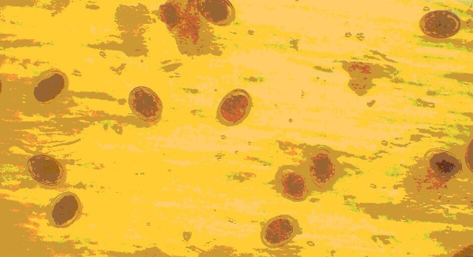
Only a microscopic examination of the feces will help determine the presence of roundworm eggs. The egg is a yellow formation with a shell covered with tubercles. Sometimes an embryo is visible in a fertilized egg. They are very resistant to environmental influences and can exist outside the human body for years.
Ascaris eggs
Since it is very difficult to detect the effects of the presence of roundworms in the body, you should be aware of the symptoms of invasion: a sudden increase in body temperature;
- skin rash;
- choking and coughing (sometimes with blood);
- muscle cramp;
- joint pain
This manifestation is associated with the effects of roundworm allergens on the body. If such symptoms are detected, it is necessary to take a stool test for worm eggs.
Where to go if you suspect worms?
If you suspect a helminthic attack, you must make an appointment with an infectious disease specialist. In the early stages, helminthiasis does not have specific symptoms, so it is quite difficult to suspect that you or a loved one has worms. As a rule, patients complain of mild pain: indigestion, headache, apathy.
If the symptoms do not disappear within a week or the condition returns periodically (for example, once every 3-4 months you feel unwell), you should see your doctor. Bad health attacks may be associated with parasite migration.
Pinworm infection
Pinworms can be contracted through casual contact with sick people (through shared objects, shaking hands). People often get attacks from cats and dogs; Worm eggs live on pet fur. Children are especially vulnerable to this disease. A child can be infected with this parasite in kindergarten or from animals. Pinworm eggs can be found on any object that has been in contact with the patient. It can be found under nails, on toys, bedding and underwear. Therefore, it is very easy to get infected with pinworms.
Pinworm eggs
Pinworms lead to the development of a disease called enterobiasis. Signs of an attack are as follows:
- itching in the area of the rectal outlet;
- diarrhea;
- nausea;
- sudden weight loss;
- bloated stomach.
Pinworm eggs are not excreted in the stool. Parasites breed in the anal area, where they lay eggs, which cause itching. To detect the presence of these worms in the body, scrapings are made from the skin of the anus and microscopic examination of the material taken is carried out. Such an analysis is usually required when children are enrolled in kindergarten. The scraping is taken in the morning before washing the child, so as not to wash the parasite eggs. Perform triplicate analysis over several days. Pinworm eggs under the microscope look like oval-shaped white grain particles.
Adult pinworms can be found in the feces of children and adults. This is a small white worm about 0. 5-1 cm long, one end of the body is pointed.
Folk remedies for helminths
For diphyllobothriasis, folk remedies should be used only after consulting a doctor. They should not replace drug treatment, but can only complement it. The most commonly used recipe is with pumpkin seeds.
Pumpkin seeds have an adverse effect on many helminths, including tapeworms. They contain cucurbitin, a substance that destroys parasites. The seeds are ground with a coffee grinder or blender, then diluted with water to a paste. For adults, you need 300 g of seeds, and for children - from 50 to 100 g. The prepared product is eaten in the morning on an empty stomach for 1 hour. After this you can not have breakfast. After 3 hours you need to take a laxative, and after another 30 minutes do an enema.
When the parasite comes out in the stool, it needs to be checked. You should pay attention to whether there is a head at one end of the body. If it is not there, then this means that only the segment has come out, and the parasite will be able to regrow the body and release the eggs. In this case, the course of treatment must be repeated.
whipworm
This type of parasite is quite rare in the central zone of our country. Whipworms often live in southern areas, because the eggs of these worms like warmth. Most infections are observed in rural areas.
Whipworm eggs live in the soil. Attacks occur through hands, contaminated soil particles, and poorly washed vegetables and fruits.
As a result of infection, a disease occurs - trichocephalosis. Whipworms parasitize the intestines. These worms cause anemia, because they feed on human blood, and severe stomach pain.
Whipworm eggs
Parasite eggs are excreted in the feces, but they are very small and cannot always be seen even under a microscope. Only with very severe infestations is it possible to detect eggs in a stool test. They are shaped like barrels and have a brownish yellow color. There are holes on both sides of the egg.
What do worms look like in stool? They are very difficult to detect living in feces, because whipworms cannot live long outside the human body. Only with anthelmintic therapy can you see dead white worms in the stool.
To diagnose trichuriasis, the rectum and sigmoid colon are examined with a special tool (sigmoidoscopy). In this way, the accumulation of parasites in the intestine is detected. Treating an attack takes a long time, because whipworm eggs are protected by a dense shell.
Diagnosis of helminthiases
When diagnosing many helminth infections, stool examination is performed first. If you find black dots in your stool or white worms in your stool, this test should be done as soon as possible.
However, not only stool with black dots is an indication for coprogram. Often, even eggs that cannot be seen by the naked eye can be easily identified under a microscope. A more accurate stool diagnosis by detecting helminth DNA particles is done using the PCR technique.
If a person has a lot of black spots in his stool, then other diagnostic methods include the following:
- Scraping from the area near the anus;
- Blood tests using ELISA, PCR, RNGA and other methods;
- Be sure to do blood biochemistry and CBC;
- To identify the localization of parasites, in some cases ultrasound, MRI and CT are performed;
- To diagnose the migration stage of helminths, X-ray examination is indicated.
For certain forms of helminthiases, examination of sputum, rectal mucus, urine, and gallbladder contents can be carried out. Endoscopic examination is also sometimes used for diagnosis.
Trichinella
This is one of the most dangerous types of roundworms. Trichinella parasitic on human muscle. Severe attacks sometimes lead to death.
Trichinella enters the body by eating poorly processed meat from wild and farmed animals. Worms are destroyed only at very high temperatures (about 80°C). Worms can be found in salted or smoked meat; such treatment does not kill their larvae.
Possible infection from undercooked meat
Parasite eggs cannot be detected in the human body. The female Trichinella carries the eggs into her body, and then the larvae are born. These are worms that reproduce ovoviviparously. It is impossible to detect Trichinella in feces. The newly born larvae immediately enter the blood and lymph, bypassing the intestines. Larvae die quickly in feces.
Usually this disease is diagnosed when the parasite has successfully entered the muscle. In this case, a person is disturbed by the following symptoms: muscle pain;
- swollen;
- febrile condition (high temperature, pain, malaise);
- irregular bowel movements with constipation or diarrhea.
To detect invasion, a blood test with a serological test is performed. This is the only method to detect Trichinella in the body.
Articles for patients with diseases diagnosed by doctors. Does not replace a doctor's appointment and should not be used for self-diagnosis.
Wide tapeworm
The human body only contains immature tapeworm eggs. They are excreted in feces and enter the external environment. With untreated wastewater, the eggs end up in the water body and begin their development there. First they end up in the bodies of freshwater crustaceans. Fish from reservoirs become infected with tapeworms when eating small crustaceans. And a person gets a helminthic attack when eating badly fried fish, infected from fresh water bodies or raw pike caviar.
Broad tapeworm eggs
Diphyllobothriasis occurs, which is indicated by the following symptoms: pain in the abdominal cavity;
- dizziness and vomiting;
- intestinal problems (constipation or diarrhea);
- loss of appetite or excessive hunger.
What does a helminth from the tapeworm class look like? This is a large parasite that can reach 10 m in length. In feces, only individual living parts (segments) of worms can be found; they look like long white ribbons (from 30 cm to 3 m). They should be removed from the feces with tweezers, transferred to a clean container and taken to a parasite or infectious disease specialist for analysis.
Microscopic examination of stool can reveal tapeworm eggs. Their size is about 0. 07 mm. The egg looks like a yellowish oval formation covered with a thick shell. One end of the egg is covered with a cap, and the other end with a bulge.
Worm larvae can be shed in the feces, but they are not dangerous. Diphyllobothriasis cannot be contracted from infected people or animals. Attacks occur exclusively through the consumption of fish.
Harm the body
When tapeworms enter the intestine, diphyllobothriasis develops. Helminths mainly affect the gastrointestinal tract. Inflammation and ulcers form on the intestinal wall where the worms attach. If there is not one, but several parasites in the body, they can clog the intestinal lumen, resulting in an obstruction. Helminths constantly irritate the walls of the gastrointestinal tract, which leads to disturbances in the digestive process. In addition, it poisons the human body with waste, which causes allergies. When the parasite remains in the body for a long time, severe anemia and vitamin B12 deficiency develop.
Cattle and pig tapeworms
People become infected with this type of parasite by eating unprocessed meat from domestic animals. Worm segments are excreted in the patient's stool. In the external environment, the segments move through the soil and lay eggs with larvae inside. These eggs are then eaten by domestic animals. When a person eats contaminated beef or pork, they become infected with beef or pork tapeworms. To kill tapeworms, you need to boil or fry the meat for at least 30 minutes.
Bull tapeworm
Cattle tapeworms cause taeniahrynchiasis, and pig tapeworms cause taeniasis. The symptoms of this disease are similar: abdominal pain;
- constant hunger;
- dizziness and vomiting;
- weakness;
- weight reduction;
- diarrhea;
- itching in the anal area when the segment comes out.
Worms in the patient's feces are in the form of segments. They look like light strips about 1-2 cm long. Pig tapeworm segments are longer and consist of 3 segments.
When analyzing feces, tapeworm eggs (oncospheres) are detected. They are round formations with a dense shell, inside which there is an embryo.
Infection with pork tapeworm is possible through dirty hands, without an intermediate host. Segments excreted in the patient's stool are dangerous. They can enter the human body from contaminated soil. In this case, pig tapeworm larvae multiply in the human body and cause a serious disease - cysticercosis. This is a very dangerous invasion. Larvae enter the brain, spinal cord, eyes, heart and lungs, causing severe damage. With cysticercosis, segments and eggs are not excreted in the stool. This disease can only be detected through serological blood tests and cerebrospinal fluid analysis.
Classification
Modern medicine classifies worms that parasitize the human body as follows: Luminal. Such worms live in the intestinal lumen. These include broad tapeworms, dwarf and bull tapeworms, hookworms, pinworms, whipworms, roundworms, etc.
Fabric. Such worms choose muscle tissue and lungs, as well as organs such as the pancreas, liver, brain, etc. for their habitat.
Depending on the exact place where the tissue helminth is localized, the invasion may have the following names:
- Filariasis. The parasite lives in the lymph nodes
- Cysticercosis. Areas of the brain affected by helminths
- Echinococcosis. Helminthic attack is diagnosed in the liver
- Paragonimiasis. The parasite lives in the lungs
Flukes
Of the flukes class of worms, the cat fluke (liver fluke) is most often found in humans. The habitat of worm eggs is fresh water. From there, the parasite enters the body of the shellfish and then into the fish. Cats and humans become infected with fluke by eating poorly processed freshwater fish, as well as through contaminated water. A sick cat does not pose a danger to humans.
Liver burbot with parasites
Most often, fish from the carp family are infected. Salting or smoking does not lead to the death of the parasite. A relatively long heat treatment of the product is required. You can get fluke by accidentally swallowing water from a pond or river. There are known cases of invasion after watering the beds with contaminated water.
Cat fluke attacks the liver. There is pain in the abdominal cavity on the right side, nausea, vomiting, fever. During the medical examination, organ enlargement was detected.
Adult worms are not excreted in the feces. What do fluke worm eggs look like under a microscope? When examining the feces, you can see a transparent oval with a golden shell. On one side of the egg there is a plug that opens when the larvae hatch. For diagnostic purposes, blood tests are also performed for antibodies or enzyme-linked immunosorbent assays.
How to know if there are worms?
It is impossible to independently determine the presence of helminthic infestation. In the early stages, the disease can be almost symptomless. The patient does not experience pain; The immune system can suppress the effects of pathogens, toxins and allergens for some time. As a rule, deterioration begins during the period of larval migration or with an increase in the number of worms. The stronger the infestation (that is, the more parasites), the more symptoms appear.
However, traveling without symptoms of invasion is dangerous - the patient infects others, and his health gradually worsens. To detect the disease, it is necessary to undergo periodic preventive examinations in the hospital. As part of prevention, the therapist prescribes a test for worms at least once a year. If you live in an endemic area - once every six months.
What can be seen with the naked eye?
Since some parasites are very small in size, it is not possible in all cases to detect their presence in the body only by the presence of eggs in the feces. Some parasites are microscopic in size and live hidden in the body, without betraying their presence. In addition, they are not always localized in the intestine and can migrate throughout the body. Therefore, to diagnose parasitic infections, they use serological tests, which are based on the antigen-antibody immune response.
All parasites look different, have their own specific development cycle, different attack symptoms and differences in treatment regimens. However, there are some symptoms that may indicate a parasitic infection in a person:
- rapid weight loss;
- intestinal disorders: diarrhea replaces constipation;
- strong itching in the anus;
- skin rash of unknown etiology;
- stomachache;
- bloated stomach;
- loss of appetite;
- inexplicable desire for sweets;
- sometimes uncontrolled appetite in adults;
- frequent colds due to a decrease in the body's defenses.